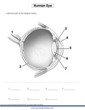
42 eye diagram with labels and functions
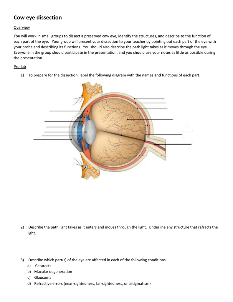
Eye Diagram With Labels and detailed description A brief description of the eye along with a well-labelled diagram is given below for reference. Well-Labelled Diagram of Eye The anterior chamber of the eye is the space between the cornea and the iris and is filled with a lubricating fluid, aqueous humour. The vascular layer of the eye, known as the choroid contains the connective tissue. Diagram of the Eye - Lions Eye Institute In order for the eye to work at its best, all parts must work well collectively. To understand the eye and its functions, it's important to understand how the eye works, see below diagrams for both the external eye and the internal eye. The External Eye Instructions Click the parts of the eye to see a description for each.
Labeled Eye Diagram | Eye anatomy diagram, Eye anatomy ... accessory structures of the eye, extrinsic eye muscles, anatomy of the eyeball and microscopic anatomy of the retina. The skeletal system consists of bones and their associated connective tissues, including cartilage, tendons, and ligaments. It consists of dynamic, living tissues that are capable of growth, detect pain stimuli, adapt to stress ...

Eye diagram with labels and functions
Eye Diagram - Labelled Diagram of Human Eye, Explanation ... The basic functions of Rods and Cones are conscious light perception, color differentiation and depth perception. The human eye is capable of distinguishing between about 10 million colors, and it can also detect a single photo. The human eye is a part of the sensory nervous system. Labeled Diagram of Human Eye Parts of the Eye & Their Function | Robertson Optical and ... Eye Parts Description and Functions; Cornea: The cornea is the outer covering of the eye. This dome-shaped layer protects your eye from elements that could cause damage to the inner parts of the eye. There are several layers of the cornea, creating a tough layer that provides additional protection. These layers regenerate very quickly, helping ... Eye anatomy: A closer look at the parts of the eye The iris of the eye functions like the diaphragm of a camera, controlling the amount of light reaching the back of the eye by automatically adjusting the size of the pupil (aperture). The eye's crystalline lens is located directly behind the pupil and further focuses light.
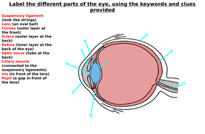
Eye diagram with labels and functions. Label Parts of the Human Eye - University of Dayton Label Parts of the Human Eye. Select One Anterior Chamber Ciliary Body Cornea Fibrous Tunic Iris Lateral Rectus Muscle Lens Medial Rectus Muscle Optic Disk Optic Nerve Pupil Retina Vascular Tunic Vitreous Nerve. Eye Anatomy: 16 Parts of the Eye & Their Functions The following are parts of the human eyes and their functions: 1. Conjunctiva The conjunctiva is the membrane covering the sclera (white portion of your eye). The conjunctiva also covers the interior of your eyelids. Conjunctivitis, often known as pink eye, occurs when this thin membrane becomes inflamed or swollen. Human Eye: Structure of Human Eye (With Diagram) | Biology The human eye is a very sensitive and delicate organ suspended in the eye socket which protects it from injuries. It essentially consists of CORNEA, LENS & RETINA besides many other parts such as Iris, Pupil and aqueous humour, vituous humour etc. Each one has got a specific function. A section of the eye is as shown in Fig. 2.2. Eye Parts Labeling and Functions Flashcards - Quizlet Start studying Eye Parts Labeling and Functions. Learn vocabulary, terms, and more with flashcards, games, and other study tools.
Eye pattern - Wikipedia In telecommunication, an eye pattern, also known as an eye diagram, is an oscilloscope display in which a digital signal from a receiver is repetitively sampled and applied to the vertical input, while the data rate is used to trigger the horizontal sweep. It is so called because, for several types of coding, the pattern looks like a series of eyes between a pair of rails. Eye Diagram - Differentiated Worksheets and ... - Pinterest Eye Diagram - Differentiated Worksheets and EASEL Activities Description Use these simple eye diagrams to help students learn about the human eye. Three differentiated worksheets are included: 1. Write the words using a word bank 2. Cut and paste the words 3. Eye Anatomy: Parts of the Eye and How We See - American ... The eye sits in a protective bony socket called the orbit. Six extraocular muscles in the orbit are attached to the eye. These muscles move the eye up and down, side to side, and rotate the eye. The extraocular muscles are attached to the white part of the eye called the sclera. This is a strong layer of tissue that covers nearly the entire ... Structure And Function Of The Eye - Vision - MCAT Content Structure and Function of the Eye The human eye is an organ that reacts with light and allows light perception, color vision, and depth perception. The photoreceptive cells of the eye, where transduction of light to nervous impulses occurs, are located in the retina (shown in Figure 1) on the inner surface of the back of the eye.
Labelling the eye - Science Learning Hub Labelling the eye Add to collection The human eye contains structures that allow it to perceive light, movement and colour differences. In this activity, students use online or paper resources to identity and label the main parts of the human eye. By the end of this activity, students should be able to: identify the main parts of the human eye Generate eye diagram - MATLAB eyediagram eyediagram (x,n,period) sets the labels on the horizontal axis to the range between - period /2 to period /2. eyediagram (x,n,period,offset) specifies the offset for the eye diagram. The function assumes that the ( offset + 1)th value of the signal and every n th value thereafter, occur at times that are integer multiples of period. Eye Anatomy | Definition, Structure & Functions Functions of Eyes are given below: Our eyes detect light. The two eyes are positioned so that each one gives a slightly different view of your surroundings. This gives us a Binocular Vision. Each eye produces an image, or picture, of what you are looking at. Information about the image in each eye is then sent along a nerve to your brain. Structure and Functions of Human Eye with labelled Diagram Structure and Functions of Human Eye with labelled Diagram Biology Biology Article Structure Of Eye Structure of the Eye The eye is one of the sensory organs of the body. In this article, we shall explore the anatomy of the eye The structure of the eye is an important topic to understand as it one of the important sensory organs in the human body.
Structure and functions of the eye by sgreen2 - Teaching Resources - Tes
Eye Anatomy Diagram - EnchantedLearning.com Retina - light-sensitive tissue that lines the back of the eye. It contains millions of photoreceptors (rods and cones) that convert light rays into electrical impulses that are relayed to the brain via the optic nerve. Rods - cells the in the retina that sense brightness (they are photoreceptors). Night vision involves mostly rods (not cones).
Free Brain Diagram, Download Free Brain Diagram png images, Free ClipArts on Clipart Library
PDF Parts of the Eye - National Eye Institute Parts of the Eye . To understand eye problems, it helps to know the different parts that make up the eye and the functions of these parts. Here are descriptions of some of the main parts of the eye: Cornea: The cornea is the clear outer part of the eye's focusing system ... Eye Diagram Handout Author:

May I have a simplest diagram of an eye, Please - Science - The Human Eye and the Colourful ...
Human Eye Diagram, How The Eye Work -15 Amazing Facts of Eye First, light rays enter the eye through the cornea, the clear front "window" of the eye. The dome shaped cornea bends light to help the eye focus. From the cornea, the light passes through an opening called the pupil. The amount of light passing through is controlled by the iris, or the colored part of your eye.

Diagram of the Ear
Anatomy of the eye: Quizzes and diagrams | Kenhub Take a look at the diagram of the eyeball above. Here you can see all of the main structures in this area. Spend some time reviewing the name and location of each one, then try to label the eye yourself - without peeking! - using the eye diagram (blank) below. Unlabeled diagram of the eye. Click below to download our free unlabeled diagram of ...
Unlabeled Eye Diagram - Cliparts.co
PDF Eye Anatomy Handout - National Eye Institute of light entering the eye. Lens: The lens is a clear part of the eye behind the iris that helps to focus light, or an image, on the retina. Macula: The macula is the small, sensitive area of the retina that gives central vision. It is located in the center of the retina. Optic nerve: The optic nerve is the largest sensory nerve of the eye.

Blank Eye Diagram - Cliparts.co
Labelling the eye - Science Learning Hub In this interactive, you can label parts of the human eye. Use your mouse or finger to hover over a box to highlight the part to be named. Drag and drop the text labels onto the boxes next to the eye diagram If you want to redo an answer, click on the box and the answer will go back to the top so you can move it to another box.
31 Label The Eye Quiz - Best Labeling Ideas
The Eyes (Human Anatomy): Diagram, Function, Definition ... One eye sees better than the other, so your brain favors that eye. The weaker eye, which may or may not wander, is called the "lazy eye." Astigmatism: A problem with the curve of your cornea. If ...
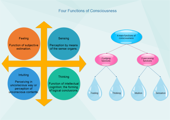
Consciousness Functions Diagram | Free Consciousness Functions Diagram Templates
Eye anatomy and function - AboutKidsHealth For people with normally functioning eyes, the following sequence takes place: Light reflects off the object we are looking at. Light rays enter the eye through the cornea at the front of the eye. The light passes through a watery fluid (aqueous humor), and enters the pupil to reach the lens.
The Eye Diagram Label Worksheets (Differentiated) by zmzb | Teaching Resources
Microscope Types (with labeled diagrams) and Functions Simple microscope labeled diagram Simple microscope functions It is used in industrial applications like: Watchmakers to assemble watches Cloth industry to count the number of threads or fibers in a cloth Jewelers to examine the finer parts of jewelry Miniature artists to examine and build their work Also used to inspect finer details on products

Male Anatomy Diagram Unlabeled : Sc 912 L 16 13 Reproductive System - Blank ear diagram human ...
The Eye Diagram: What is it and why is it used? The eye diagram is used primarily to look at digital signals for the purpose of recognizing the effects of distortion and finding its source. To demonstrate using a Tektronix MDO3104 oscilloscope, we connect the AFG output on the back panel to an analog input channel on the front panel and press AFG so a sine wave displays. Then we press Acquire.

Blank Ear Diagram | Human ear diagram, Ear anatomy, Ear diagram
Eye anatomy: A closer look at the parts of the eye The iris of the eye functions like the diaphragm of a camera, controlling the amount of light reaching the back of the eye by automatically adjusting the size of the pupil (aperture). The eye's crystalline lens is located directly behind the pupil and further focuses light.

Labeled Eye Diagram - ClipArt Best
Parts of the Eye & Their Function | Robertson Optical and ... Eye Parts Description and Functions; Cornea: The cornea is the outer covering of the eye. This dome-shaped layer protects your eye from elements that could cause damage to the inner parts of the eye. There are several layers of the cornea, creating a tough layer that provides additional protection. These layers regenerate very quickly, helping ...

Module 1: Labeled Diagram of the Eye | Eye health | Pinterest | Activities
Eye Diagram - Labelled Diagram of Human Eye, Explanation ... The basic functions of Rods and Cones are conscious light perception, color differentiation and depth perception. The human eye is capable of distinguishing between about 10 million colors, and it can also detect a single photo. The human eye is a part of the sensory nervous system. Labeled Diagram of Human Eye
Labeled Eye Diagram - ClipArt Best
Eye Diagram With Labels And Functions - Aflam-Neeeak

Microscope Parts and Functions

draw a diagram of the human eye as seen in a vertical section and label the part which suits the ...
Post a Comment for "42 eye diagram with labels and functions"