40 onion cells under microscope with labels
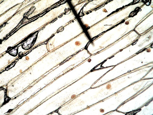
Under the Micrsocope: Onion Cell (100x - 400x) - YouTube In this "experiment" we will see onion cells under the microscope.For the experiment you will only need onion, dropper and the microscope (container and tool... The Cell Structure of an Onion | Sciencing Cell Walls Give Structure. Cell walls in plants are rigid, compared to other organisms. The cellulose present in the cell walls forms clearly defined tiles. In onion cells the tiles look very similar to rectangular bricks laid in offset runs. The rigid walls combined with water pressure within a cell provide strength and rigidity, giving plants ...
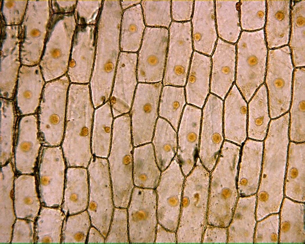
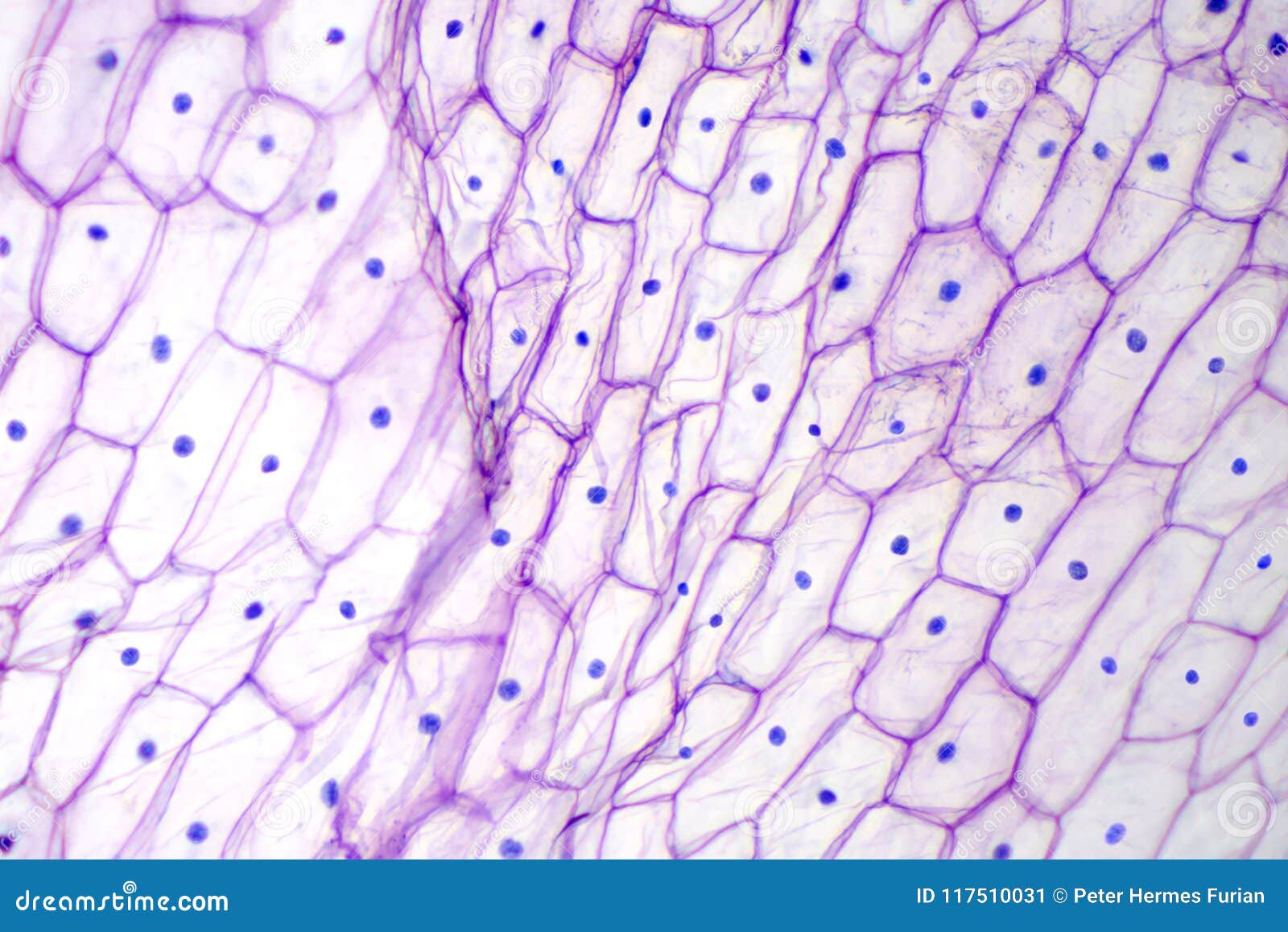
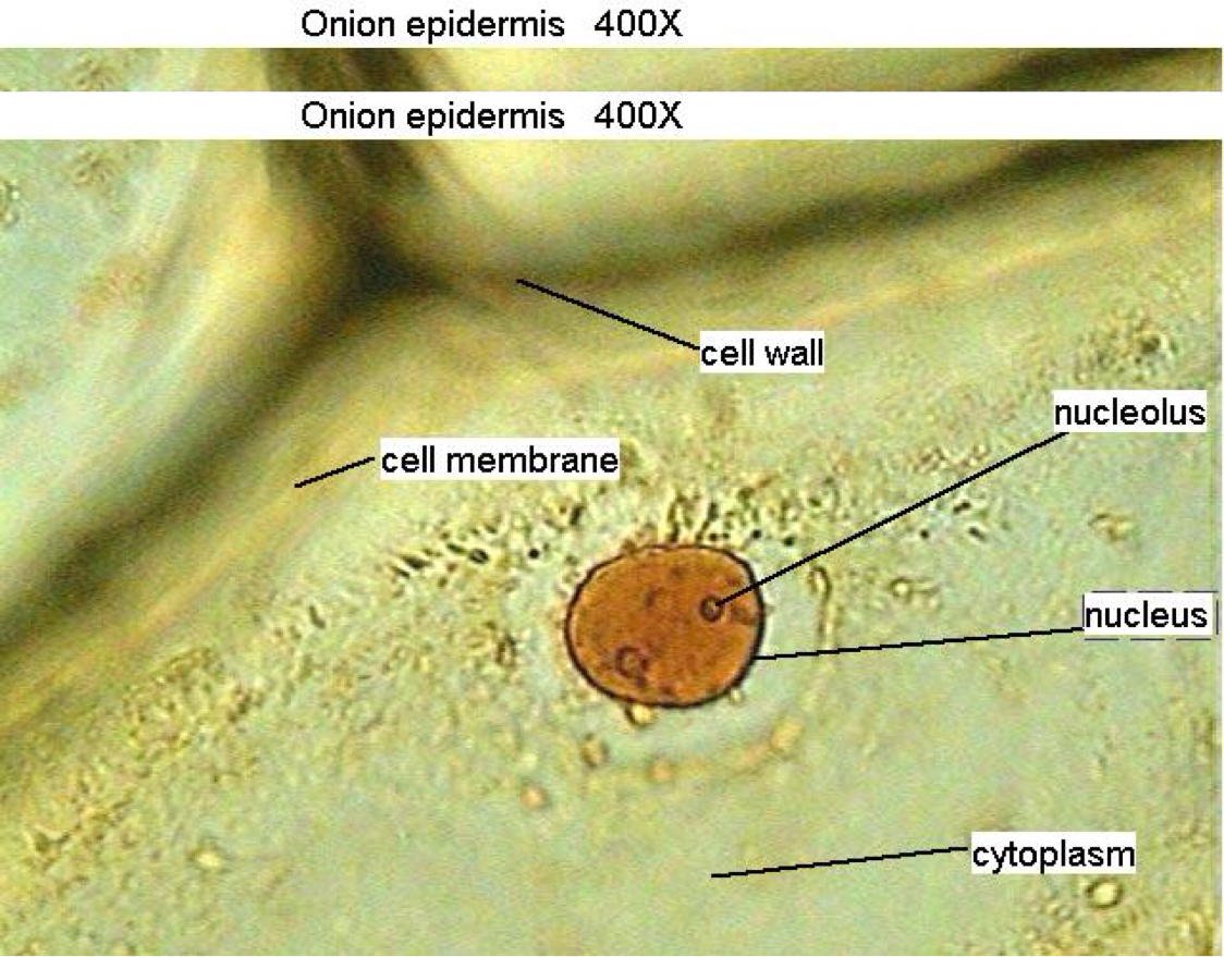
onion cells under a microscope labeled Onion epidermal cells, iodine stain, 400X. distichum 'Spence' (C and D), and sorghum cv BTx623 (E and F) grown in freshwater were imaged under a light microscope Location Tiruvanmiyur, Chennai 600041 Financial Sentiment Analysis Github Label an de diagram of a stomatal apparatus 2 four parts on the diagram Inference The cells observed under the ...

Onion cells under microscope with labels
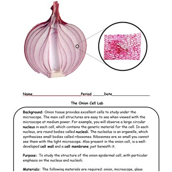
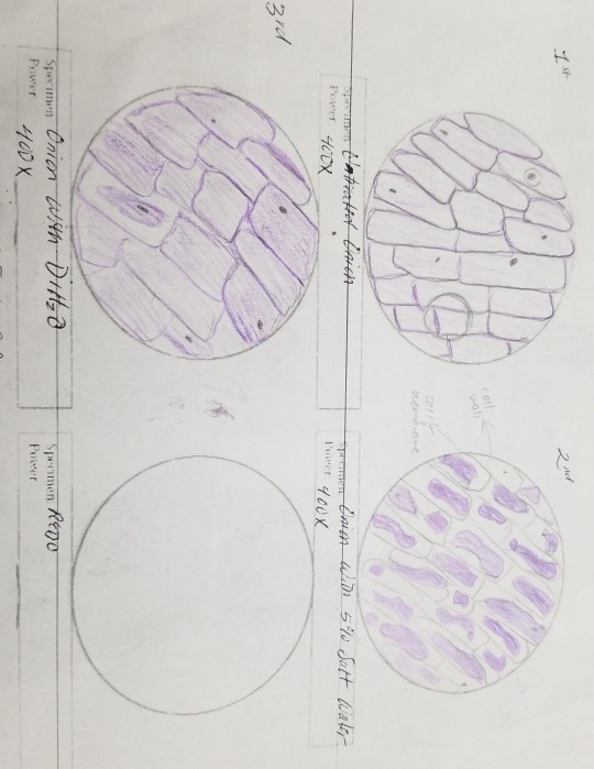
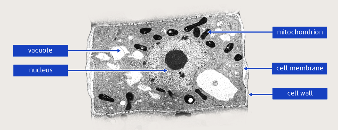
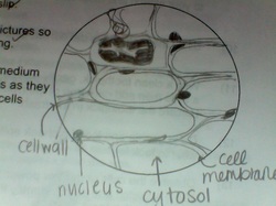
Onion Cells Under a Microscope - Requirements/Preparation/Observation Having observed the onion cell under the microscope, students will be able to learn the differences between animal and plant cells in addition to the function of the different parts of the cell. Learn about Onion Root Tip Mitosis. View Epidermal Cells. Check out other microscope experiment viewing Cheek Cells, Cork Cells or Sugar Crystals as ... What organelles are in an onion cell? - Biology Stack Exchange You cannot see most of these as they appear translucent as well as being too small to see under the light microscope. You need an electron microscope to view these. ... chloroplasts are not present in an onion cell as it is not a photosynthesising cell. This is a typical onion cell slide with labels: Share. Improve this answer. Follow edited ... Microscope Lab - SKIN AND ONION CELLS.pdf - Course Hero Observe the onion cell under both low and high power. Make a drawing of one onion cell, labeling all of its parts as you observe them. (At minimum you should observe the nucleus, cell wall, and cytoplasm.) 4. Draw and label the onion cell at both low and high power: Procedure: Skin cells - each person will make their own slide 1.
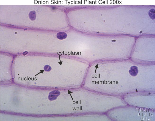
Onion cells under microscope with labels. Onion Plant Cell Under Microscope Labeled / Onion Cells - Onion ... Cell Under Microscope High Res Stock Images Shutterstock from image.shutterstock.com Draw and label the onion cell at both low and high power: Clear epidermal cells of an onion, allium cepa, in a single layer. Faites votre choix parmi les nombreuses scènes similaires. Microscope Cell Lab: Cheek, Onion, Zebrina | SchoolWorkHelper Microscope Cell Lab: Cheek, Onion,…. Introduction. The purpose of this lab was to use the microscope and identify cells such as animal cells and plant cells. This subject is important because in Biology, we will be using the microscope many times during different laboratory exercises. The microscope is used for looking at many specimens that ... PDF Onion Cell Lab Research Biology Onion Cell Lab page 1 of 3 Onion Cell Lab After you have completed the rest of this lab come back to this cover page DRAW & LABEL AN ONION CELL WITH ALL THE PARTS / ORGANELLES YOU OBSERVE UNDER 40X. Purpose: To observe and identify major plant cell structures and to relate the structure of the cell to its function. Materials: 1 ... Onion Cells Under Microscope With Labels - Realtec Find and download Onion Cells Under Microscope With Labels image, wallpaper and background for your Iphone, Android or PC Desktop. Realtec have about 34 image published on this page. onion microscope under cells cepa allium slide footage shutterstock background royalty. Pin It.
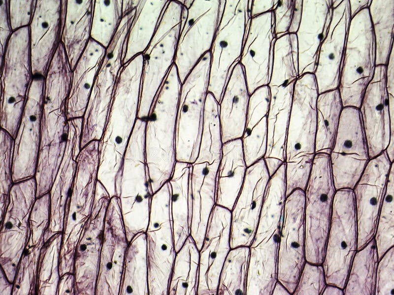
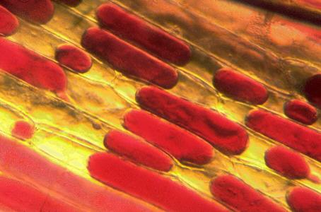
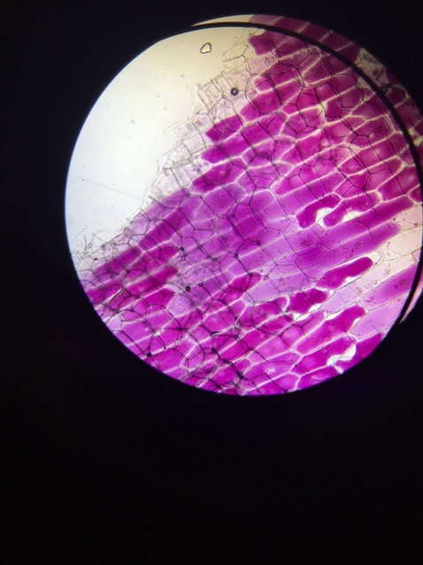
What do you observe onion cells under a microscope? Staining Onion Cells. Since onion peels are translucent, you'll need to stain the onion cells before you observe them under the microscope. There are different types of stains depending on what type of cell you are going to look at. Iodine- dark stain that colors starches in cells. In an onion cell, it will make the cell wall more visible. onion cells under a microscope labeled - sevadham.net onion cells under a microscope labeled. dream team 1992 training. onion cells under a microscope labeledprogramming in scala github. black white and cream living room ideas ... Under Onion Cell Microscope Labeled [429ORE] Label the nucleus, cell membrane, and cytoplasm 6 A drawing of one of the cells as seen under high power is shown below Allow the iodine to soak into the onion for two minutes before observing the cell under the microscope Exercise 3A some classic onion cells under my microscope ( lomo biolam ) at 56x and 315x hope you enjoy some classic onion ... Observing Onion Cells Under The Microscope » Microscope Club Summary. Observing onion cells under a microscope is a fun and easy activity for students and hobbyists alike. Onion epidermal cells appear as a single thin layer and look highly organized and structured in terms of shape and size. Certain parts of the cell are also clearly distinguishable with or without staining, making the activity even ...
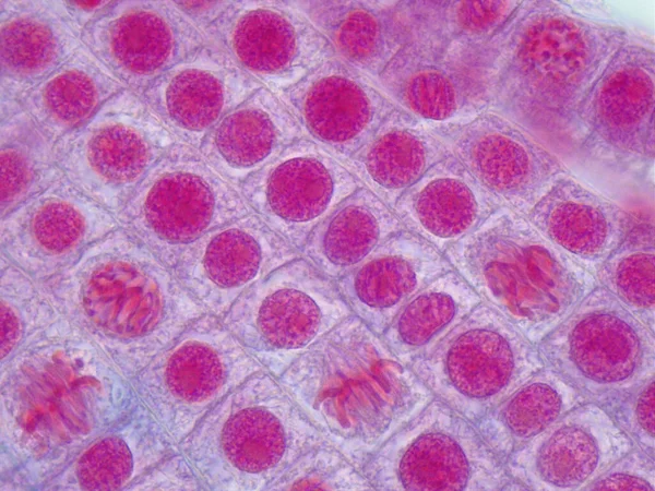
How to see onion cell with Homemade Microscope Easy - YouTube Hi friends I am Khilesh and going to show you onion cells in my homemade microscope. Tip:- Stain the cells with water then with iodine then again with water.... Onion Skin Epidermis Sample under microscope 4x,10x Magnification A sample on an onion skin epidermis diyed in blue for visibility, viewd under the microscope at 4x and 10x magnification.microscope:Biolux model :AL PDF Onion Cells - Investigation 5. Observe the onion tissue under the microscope at 4x, 10x and 40x with lots of light (open diaphragm). Then slowly close the diaphragm while observing the image to find the best light for seeing cellular details. 6. Draw a section of onion skin cells at 10x magnification. Then switch to 40x and draw one cell and label it. Questions: 1. Cell Under Labeled Microscope Onion [0FDNC6] Search: Onion Cell Under Microscope Labeled. Part C: Onion Cells Volvox are colonial flagellates and a very popular organism for classroom observations Size of onion cell-1600/2=800 µm 1cm2 is sufficient Obtain a slide of onion root cells Obtain a slide of onion root cells.
How to Observe Onion Cells under a Microscope - Blog, She Wrote
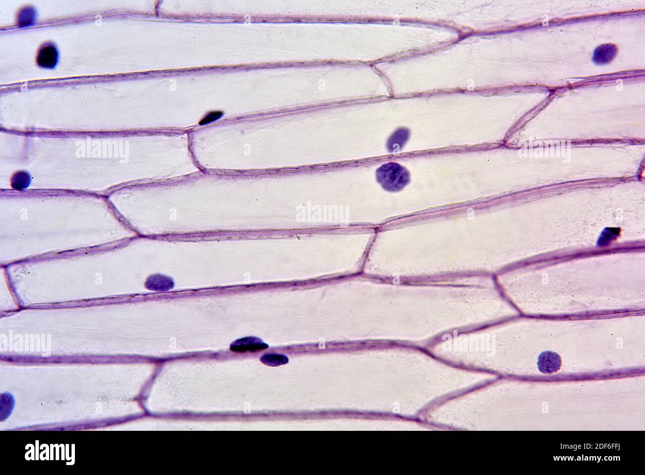
Onion Cell Lab Report.docx - Onion Cell Lab Report By Onion Cell Lab Report By : Nawaf Almalki Introduction: Many things that are viewed using a microscope, particularly cells, can appear quite transparent under the microscope. The internal parts of the cells, the organelles, are so transparent that they are often difficult to see. Biologists have developed a number of stains that help them see the cells and their organelles by adding color to ...
Onion Epidermis 100X: General Biology Lab: Loyola University ...
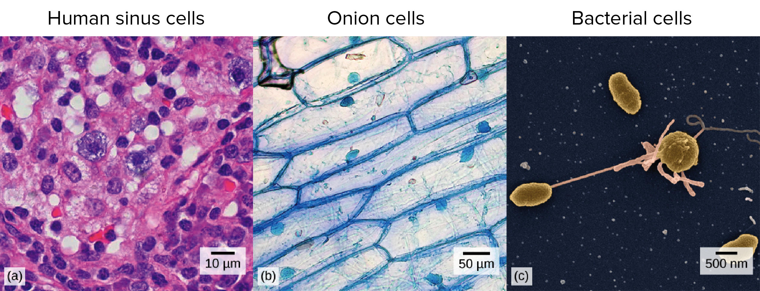
DOC Plant and Animal Cells Microscope Lab - hillsboro.k12.oh.us Students will observe onion cells under a microscope. Students will discover that their skin is made up of cells. Students will observe cheek cells under a microscope. Materials: microscope. ... Draw a diagram of one cheek cell and label the parts. (You should observe the cell membrane, nucleus, and cytoplasm.)

Of the given slide observations spot the correct sequence ...
Microscope Lab - SKIN AND ONION CELLS.pdf - Course Hero Observe the onion cell under both low and high power. Make a drawing of one onion cell, labeling all of its parts as you observe them. (At minimum you should observe the nucleus, cell wall, and cytoplasm.) 4. Draw and label the onion cell at both low and high power: Procedure: Skin cells - each person will make their own slide 1.
Onion cells microscope hi-res stock photography and images ...
What organelles are in an onion cell? - Biology Stack Exchange You cannot see most of these as they appear translucent as well as being too small to see under the light microscope. You need an electron microscope to view these. ... chloroplasts are not present in an onion cell as it is not a photosynthesising cell. This is a typical onion cell slide with labels: Share. Improve this answer. Follow edited ...

Under The Microscope Onion Cells Stock Photo, Picture And ...
Onion Cells Under a Microscope - Requirements/Preparation/Observation Having observed the onion cell under the microscope, students will be able to learn the differences between animal and plant cells in addition to the function of the different parts of the cell. Learn about Onion Root Tip Mitosis. View Epidermal Cells. Check out other microscope experiment viewing Cheek Cells, Cork Cells or Sugar Crystals as ...
Intro to cells (article) | Khan Academy
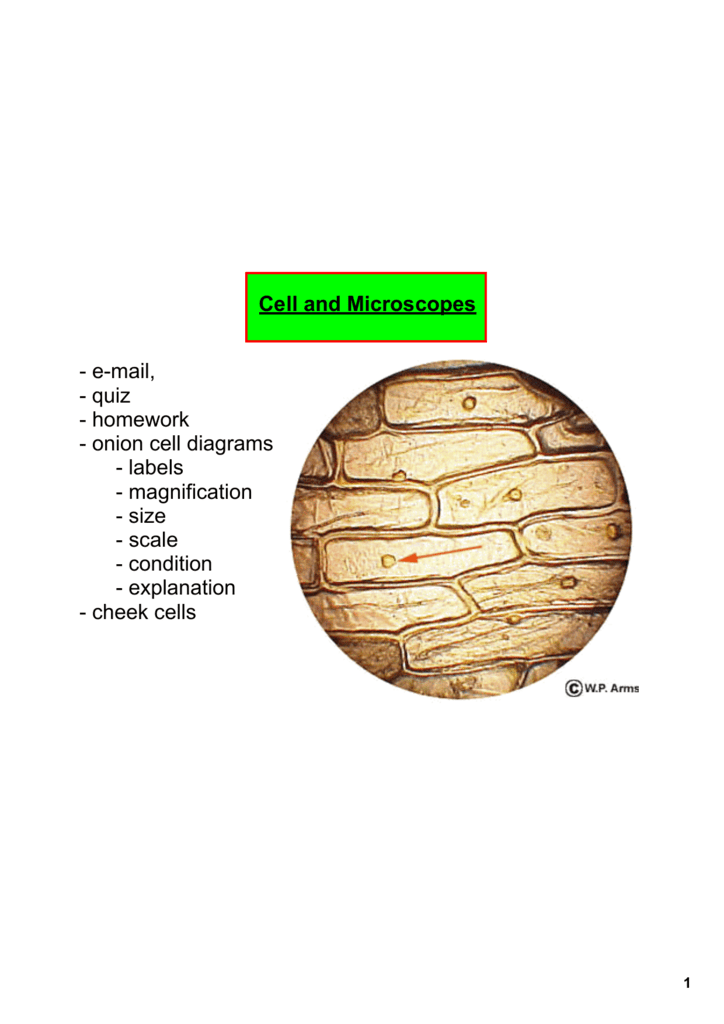
Cell and Microscopes email, quiz homework onion cell diagrams

Lab #1 microscope structure & function
STANDARD OPERATING PROCEDURE:
Onion Cell Microscope Lab

Lab Manual Exercise # 1
Onion Cell prep

Onion Cells Under the Microscope Stock Image - Image of ...
Onion cell Stock Photos, Royalty Free Onion cell Images ...

Tonicity-Onion cell lab | Miranda's AP Biology Blog
Onion skin 200x - Dissection Connection
321 Onion Cells Stock Photos - Free & Royalty-Free Stock ...

OBSERVING ONION PEEL EPIDERMAL CELLS UNDER MICROSCOPE | BEST DEMO | BIOLOGY

3,018 Onion Cells Stock Photos, Pictures & Royalty-Free ...

Cells Under A Microscope by Jaimarie Nelson
Solved I did a bio lab where I looked at purple onion skin ...
Onion Epidermis with Large Cells Under Microscope Stock Image ...

File:Onion Cells Under the Microscope.jpg - Wikimedia Commons
Mic-UK: MICROSCOPY UK / MICSCAPE - Onion epidermis, plasmolysis
Required Practical Review

Sketch the onion peel cell as seen under the microscope ...
3.3 Lesson: Cells under the microscope - Stile
Why does knowing onion cells relate to everyday life? - Quora

Cell structure Learning Intention: - ppt video online download
Biology Pictures: Onion Cells under Microscope

Structure and Function of Cells (Learn) : Biology : Class 8 ...

Sketch the onion peel cell as seen under the microscope ...
Onion cells under the microscope under two CONDITIONS ...
Cells Under A Microscope by Jaimarie Nelson
Personal Experience with Microscopes - AyushiSinhaMicroscopy

Under The Microscope Onion Cells Stock Photo, Picture And ...

Why is iodine stain used on onion cells? | Iodine, Cell, Stain
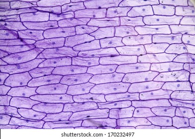
Onion Epidermis Microscopic View Stock Photo 170232497 ...
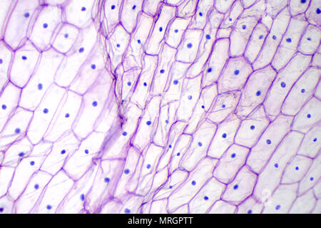
Onion cells under a light microscope at 10 times ...
What is the shape of an onion cell? Why? - Quora
Post a Comment for "40 onion cells under microscope with labels"