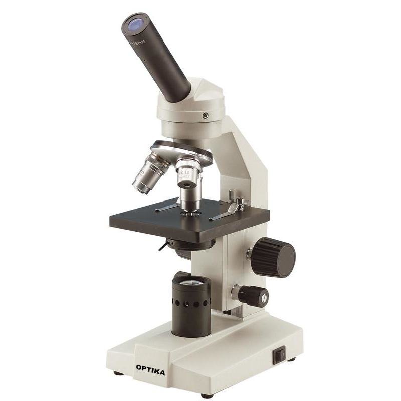
43 light microscope with labels
Principles of Birefringence | Nikon’s MicroscopyU In order to examine more closely how birefringent, anisotropic crystals interact with polarized light in an optical microscope, the properties of an individual crystal will be considered. The specimen material is a hypothetical tetragonal, birefringent crystal having an optical axis oriented in a direction that is parallel to the long axis of ... Label the Light Microscope - Labelled diagram Eyepiece, Light Source, Base, Stage, Stage Clips, Fine Focus, Coarse Focus, Arm, Objective Lens, Diaphragm.
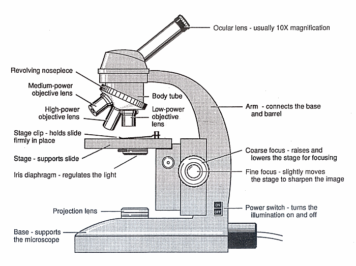
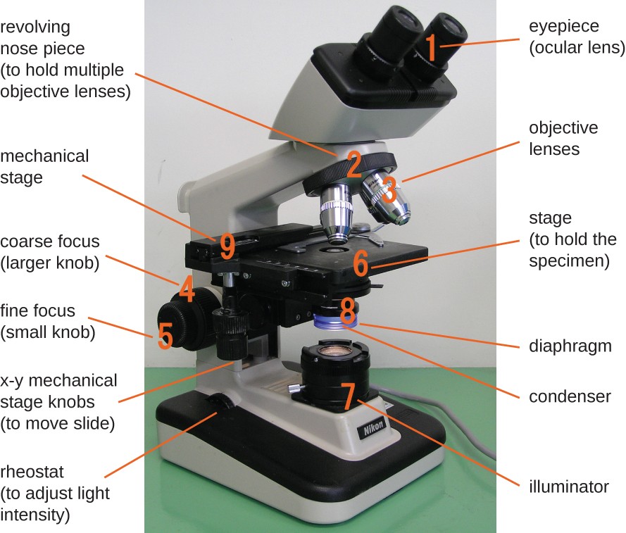
Parts of a microscope with functions and labeled diagram - Microbe Notes Q. Differentiate between a condenser and an Abbe condenser. Ans. Condensers are lenses that are used to collect and focus light from the illuminator into the specimen. They are found under the stage next to the diaphragm of the microscope. They play a major role in ensuring clear sharp images are produced with a high magnification of 400X and above.

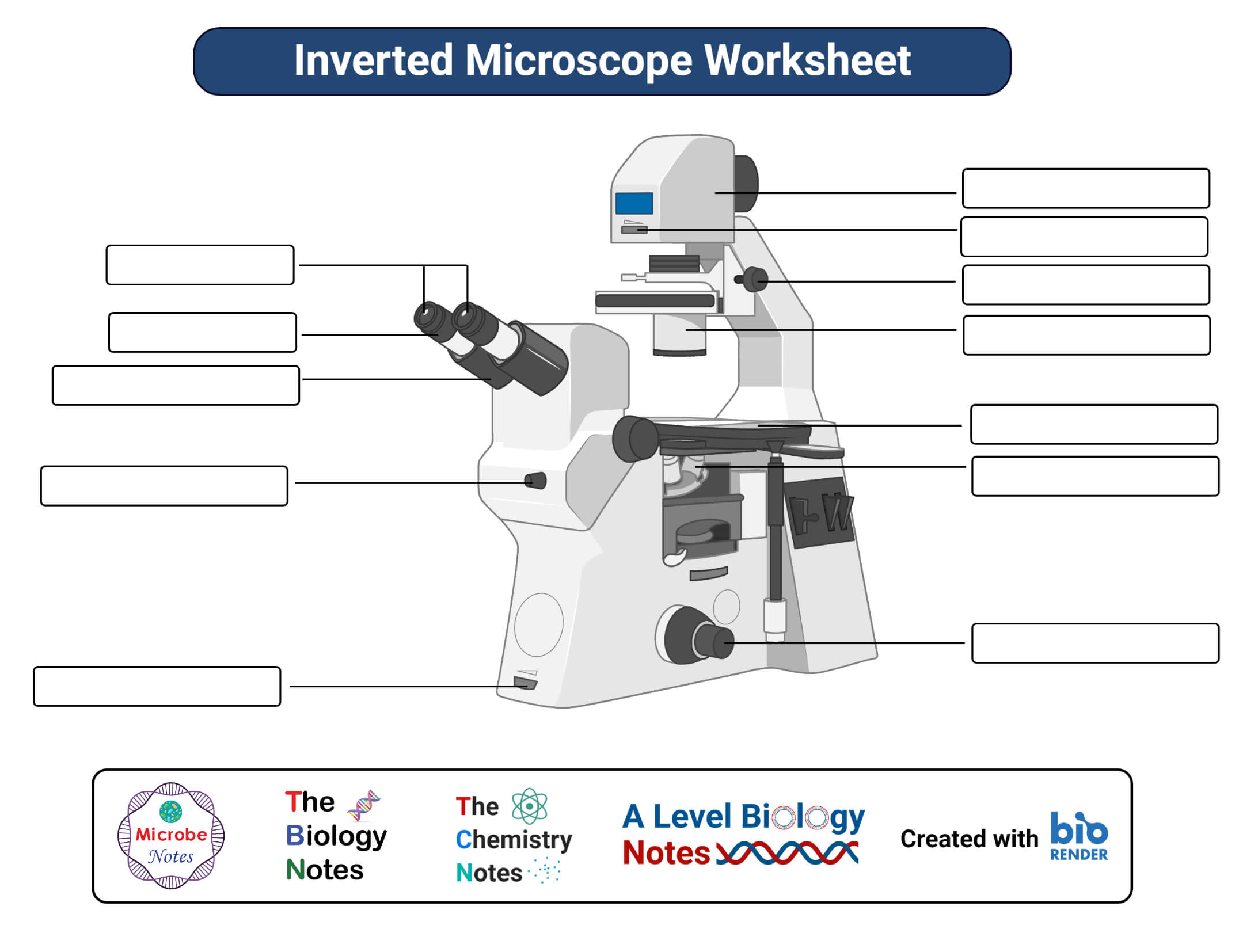
Light microscope with labels
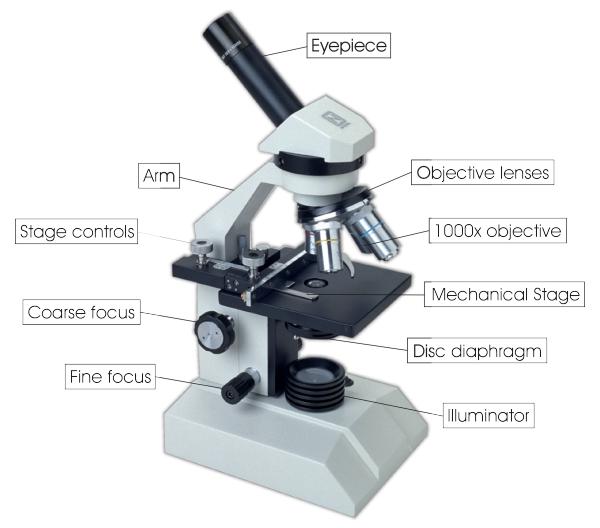
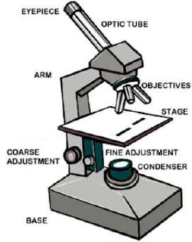
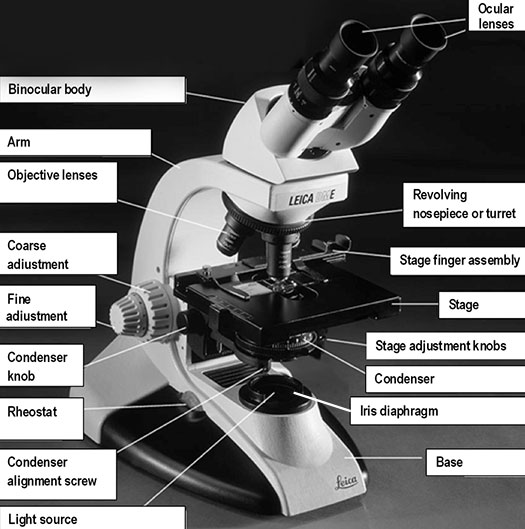
Compound Microscope Parts, Functions, and Labeled Diagram Compound Microscope Definitions for Labels. Eyepiece (ocular lens) with or without Pointer: The part that is looked through at the top of the compound microscope. Eyepieces typically have a magnification between 5x & 30x. Monocular or Binocular Head: Structural support that holds & connects the eyepieces to the objective lenses. Could Call of Duty doom the Activision Blizzard deal? - Protocol Oct 14, 2022 · “The CMA is concerned that having full control over this powerful catalogue, especially in light of Microsoft’s already strong position in gaming consoles, operating systems, and cloud infrastructure, could result in Microsoft harming consumers by impairing Sony’s — Microsoft's closest gaming rival — ability to compete,” the report ... Compound Microscope - Diagram (Parts labelled), Principle and Uses The three structural components include: 1. Head - This is the upper part of the microscope that houses the optical parts 2. Arm - This part connects the head with the base and provides stability to the microscope. Arm is used to carry the microscope around 3. Base - Base is on which the microscope rests and the base houses the illuminator that lights up the specimens
Light microscope with labels. Microscope Parts and Functions Eyepiece: The lens the viewer looks through to see the specimen. The eyepiece usually contains a 10X or 15X power lens. Diopter Adjustment: Useful as a means to change focus on one eyepiece so as to correct for any difference in vision between your two eyes. Body tube (Head): The body tube connects the eyepiece to the objective lenses. Arm: The arm connects the body tube to the base of the ... Microscope Labeling - The Biology Corner Microscope Labeling. This simple worksheet pairs with a lesson on the light microscope, where beginning biology students learn the parts of the light microscope and the steps needed to focus a slide under high power. The labeling worksheet could be used as a quiz or as part of direct instruction where students label the microscope as you go ... Compound Light Microscope Labelling Quiz - PurposeGames.com This is an online quiz called Compound Light Microscope Labelling. There is a printable worksheet available for download here so you can take the quiz with pen and paper. Your Skills & Rank. Total Points. 0. Get started! Today's Rank--0. Today 's Points. One of us! Game Points. 15. Word to HTML - Online Converter and Cleaner - 𝗪𝗼𝗿𝗱𝗛𝗧𝗠𝗟.𝗰𝗼𝗺 Free online Word to HTML converter with code cleaning features and easy switch between the visual and source editors. It works perfectly for any document conversion, like Microsoft Word
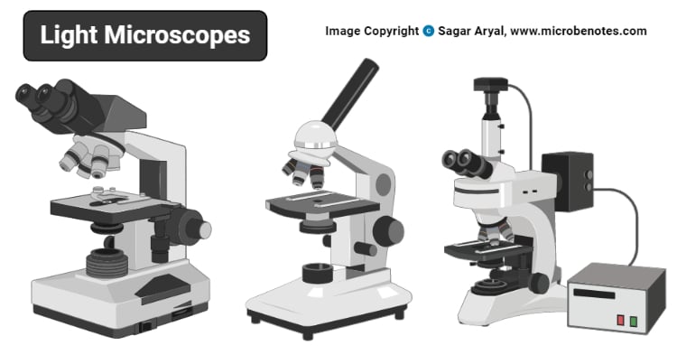
Lifestyle | Daily Life | News | The Sydney Morning Herald The latest Lifestyle | Daily Life news, tips, opinion and advice from The Sydney Morning Herald covering life and relationships, beauty, fashion, health & wellbeing Light Microscope-Definition, Principle, Types, Parts, Labeled Diagram ... The lenses are placed in such a way that they may deflect light to magnify images effectively. The ability of the light microscope to create an image by concentrating a ray of light through a small, clear object is fundamental to its operation. The picture is then magnified for viewing via one or more lenses. Label the Light Microscope - Labelled diagram - Wordwall Drag and drop the pins to their correct place on the image.. Eyepiece, Light Source, Base, Stage, Stage Clips, Fine Focus, Coarse Focus, Arm, Objective Lens. Parts of a Microscope - The Comprehensive Guide Step 1: Fully open field and condenser diaphragms and focus on specimen using x10 objective. Step 2: Fully close field diaphragm and adjust the condenser and focus so edges are as sharp as possible. Step 3: Use screws at front of condenser to centre field diaphragm and open field diaphragm to fill view. Step 4: Remove eyepiece and close down ...
Microscope Labeling Game - PurposeGames.com About this Quiz. This is an online quiz called Microscope Labeling Game. There is a printable worksheet available for download here so you can take the quiz with pen and paper. This quiz has tags. Click on the tags below to find other quizzes on the same subject. Science. Microbiology Module 1 Flashcards | Quizlet Label the terms or descriptions in the chart to assess your knowledge of the levels of the four types of macromolecules. Use the hints to help you place the labels correctly. ... - in the compound light microscope, it is the image created by the objective lens - initial image of the specimen. Microscope Label Worksheets - K12 Workbook Worksheets are The microscope parts and use, Parts of the light microscope, Label parts of the microscope, Labeling scientific tools microscope name, Parts of the microscope quiz, Use the word list to help you label the 12, Label parts of the microscope answers, Microscope lab. *Click on Open button to open and print to worksheet. Parts of the Microscope with Labeling (also Free Printouts) 5. Knobs (fine and coarse) By adjusting the knob, you can adjust the focus of the microscope. The majority of the microscope models today have the knobs mounted on the same part of the device. Image 5: The circled parts of the microscope are the fine and coarse adjustment knobs. Picture Source: bp.blogspot.com.
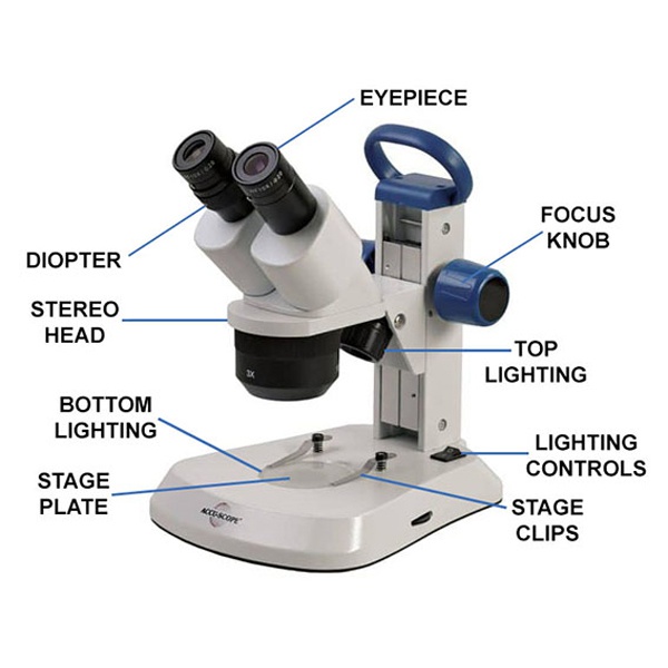
Dissecting Stereo Microscope Parts and Functions
Simple Microscope - Diagram (Parts labelled), Principle, Formula and Uses The magnification power of a simple microscope is expressed as: M = 1 + D/F. Where. M = Magnification power. D = the lease possible distance of distinct vision of eye, typically 25cm. F = Focal length of the convex lens. It is to be noted that. The magnification power of a simple microscope is about 10, meaning that the specimen being studied ...

Compound Microscope Parts, Functions, and Labeled Diagram ...
Compound Microscope- Definition, Labeled Diagram, Principle, Parts, Uses The optical microscope often referred to as the light microscope, is a type of microscope that uses visible light and a system of lenses to magnify images of small subjects. The term "compound" in compound microscopes refers to the microscope having more than one lens. Devised with a system of combination of lenses, a compound microscope ...

Compound Microscope Parts, Functions, and Labeled Diagram ...
Light microscope labels Flashcards | Quizlet Light microscope labels. Flashcards. Learn. Test. Match. Flashcards. Learn. Test. Match. Created by. school1329. Terms in this set (14) Ocular lens. First automatic magnification (x10) Body tube. Holds ocular lense. Revolving nose piece. Holds and allows selection of desired objective lens. Lowest power objective lens.
Solved PLEASE HELP THANK YOU- LIGHT SOURCE WILL BE NUMBER 1 ...
Label the microscope — Science Learning Hub All microscopes share features in common. In this interactive, you can label the different parts of a microscope. Use this with the Microscope parts activity to help students identify and label the main parts of a microscope and then describe their functions.. Drag and drop the text labels onto the microscope diagram. If you want to redo an answer, click on the box and the answer will go back ...
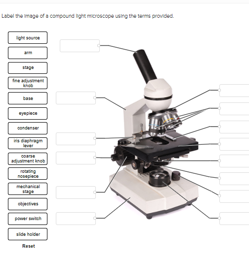
Solved Label the image of a compound light microscope using ...
Réservez des vols pas chers et trouvez des offres ... - easyJet Réservez des vols pas chers sur easyJet.com vers les plus grandes villes d'Europe. Trouvez aussi des offres spéciales sur votre hôtel, votre location de voiture et votre assurance voyage.

OMAX 40X-2500X Trinocular Biological Compound Microscope with Replaceable LED Light
Event-driven acquisition for content-enriched microscopy ... Sep 08, 2022 · Event-driven acquisition uses neural-network-based recognition of specific biological events to trigger switching between slow and fast super-resolution imaging, enriching the capture of ...

Parts of a Microscope - SmartSchool Systems
Light Microscope- Definition, Principle, Types, Parts, Labeled Diagram ... A light microscope is a biology laboratory instrument or tool, that uses visible light to detect and magnify very small objects and enlarge them. They use lenses to focus light on the specimen, magnifying it thus producing an image. The specimen is normally placed close to the microscopic lens.
Label a microscope - Teaching resources
ProSciTech Laboratory supplies and Lab equipment for Histology, Pathology, Light Microscopy, Electron Microscopy and specialist researchers.
Free Microscope, Download Free Microscope png images, Free ...
Compound Microscope Parts - Labeled Diagram and their Functions Each objective has its information (i.e. magnification) and color-code label on the side. Photo credit: Accu-scope. Usually, a compound microscope comes with 3 or 4 objective lenses. ... The illuminator is the light source for a microscope, typically located in the base of the microscope. Halogen bulbs are commonly used to provide a steady ...
Guide to using a microscope - Home
Compound Microscope - Diagram (Parts labelled), Principle and Uses The three structural components include: 1. Head - This is the upper part of the microscope that houses the optical parts 2. Arm - This part connects the head with the base and provides stability to the microscope. Arm is used to carry the microscope around 3. Base - Base is on which the microscope rests and the base houses the illuminator that lights up the specimens
Parts of a Microscope Quiz
Could Call of Duty doom the Activision Blizzard deal? - Protocol Oct 14, 2022 · “The CMA is concerned that having full control over this powerful catalogue, especially in light of Microsoft’s already strong position in gaming consoles, operating systems, and cloud infrastructure, could result in Microsoft harming consumers by impairing Sony’s — Microsoft's closest gaming rival — ability to compete,” the report ...
Instruments of Microscopy | Microbiology | | Course Hero
Compound Microscope Parts, Functions, and Labeled Diagram Compound Microscope Definitions for Labels. Eyepiece (ocular lens) with or without Pointer: The part that is looked through at the top of the compound microscope. Eyepieces typically have a magnification between 5x & 30x. Monocular or Binocular Head: Structural support that holds & connects the eyepieces to the objective lenses.
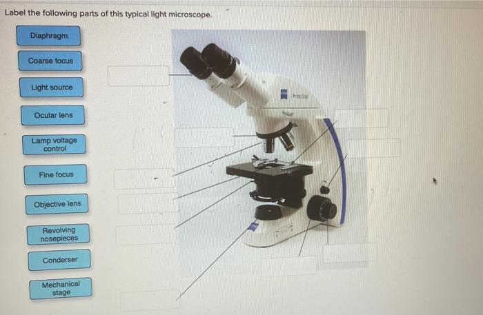
Solved Label the following parts of this typical light ...
Compound Microscope Parts, Functions, and Labeled Diagram ...

The Microscope Types of Microscopes Compound light microscope ...

Compound Microscope – Diagram (Parts labelled), Principle and ...

Monday 10/19/15 AIM: how do the parts of the compound light ...
Parts of a microscope with functions and labeled diagram

Microscope Parts and Functions
Light Microscope- Definition, Principle, Types, Parts ...
What is a Stereo Microscope? - New York Microscope Company
Microscopes

Microscope Diagram Labeled, Unlabeled and Blank | Parts of a ...

Label a Microscope Worksheet

The Science Break - Labels for the light microscope for GCSE ...

Microscope Labeling #1 Diagram | Quizlet

Photo Compound microscope with labels Image #3850568

Living Environment Course

label microscope diagram | Charts | Microscope, Anatomy bones ...

Microscope Maintenance Tips | Science supplies, Microscope ...

Label Microscope Diagram - EnchantedLearning.com

A Study of the Microscope and its Functions With a Labeled ...

microscope | Types, Parts, History, Diagram, & Facts | Britannica

Microscope With Labels Clip Art at Clker.com - vector clip ...

TYPES OF LIGHT MICROSCOPE: 1. SIMPLE... - Microbiologists_2 ...

Parts of the Microscope Label and Definition Diagram | Quizlet

Parts of a Microscope with Their Functions – Microbe Online

Compound Microscope Parts – Labeled Diagram and their ...

PARTS OF MICROSCOPE| LEARN TO LABEL COMPOUND MICROSCOPE| JUST IN 5 MINS| EXPLANATION OF PARTS
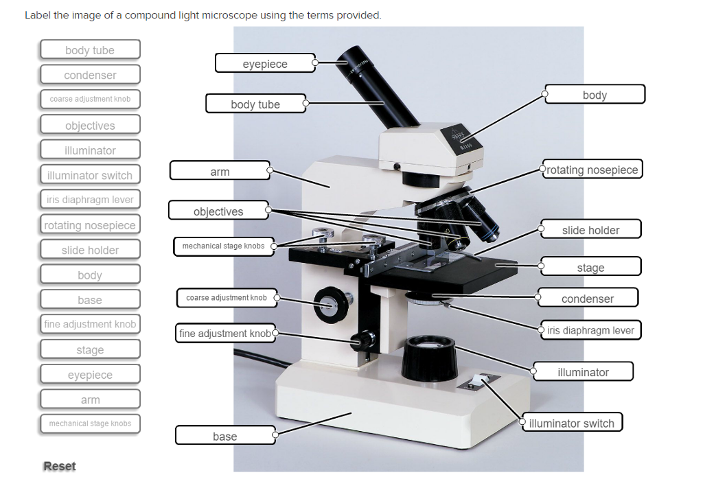
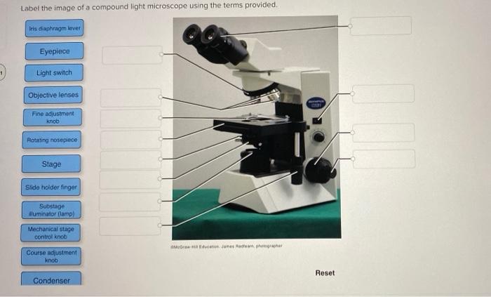
Solved Label the image of a compound light microscope using ...
The Compound Light Microscope Label the following parts on ...
Microscopes | Idaho State University
Microscope labelling - Teaching resources
Post a Comment for "43 light microscope with labels"